
the eyes are the windows to the soul but
they may be able to show you much more
than that hi I’m Rebecca Braden and
welcome to watchmojo.com and today we’ll
be seeing how a simple I exam may be
able to detect the early signs of
diabetic retinopathy so we’ll start by
having you introduce yourself my name is
dr. Ben cert I’m the director of telecom
interior of homology in the state of New
Jersey I’m responsible for all the telus
screenings so we do schools churches
runaway centers and soup kitchens how
does this exam work the test takes
roughly about 30 seconds per I we take a
color image and from the color image we
can use a computer to divide that and to
look for vision threatening disease
diabetes of course we’re looking for
glaucoma in age-related macular
degeneration then we’re going to take a
second image which is called
autofluorescence which looks
specifically at your RPE retinal pigment
epithelium and gives us a good idea on
how healthy your eye is how does
diabetes affect the eyes as we get
acquired diabetes roughly 10 years if
uncontrolled it’s going to go into the
i’ll create diabetic retinopathy when it
creates diabetic retinopathy it it
actually has the blood vessels break and
start leaking and when they leak they
actually bleed inside the eye and as
they bleed and the bleeders become
larger they actually Rob vision from the
patient what kind of lifestyle changes
can be put in place to avoid diabetic
retinopathy what we can do we have to
take charge of our own body and that
means we have to do exercise 150 minutes
a week we have to change our eating
habits we have to introduce better
lifestyle we have to stop smoking and so
on support for example if you have
diabetes and you smoke you’re going to
have cataracts ten years before anybody
else so we need to be just more in
control of what we do can you tell us
about the examiner about to see what
we’re going to do is we’re going to have
you place your your chin in a chin rest
and we’re going to bring an infrared
light towards your eye and your eye
doesn’t detect infrared we’re going to
be able to look inside your eye in your
retina and be able to capture an image
with a small flash a white flash and
that will show us what
happening and that will digitally store
the image and then we’re going to be
able to separate the images in various
colors to look at the different layers
inside the eye and see what disease we
may find then we’re going to take a
second photograph which is a little bit
brighter which will allow us to study
the RPE retinal pigment epithelium very
good that was much brighter okay very
good okay so what are we looking at here
well everything is done we’re looking at
your fondest this is the back of the art
the pattern that you see is more precise
than a fingerprint we’re looking at
arteries and we’re looking at veins and
we’re looking at these white lines those
would be nerve fiber layer so we’re
talking about glaucoma so your cup to
disc ratio is perfect your artery vein
ratio is also very good and we’re then
going to look at your macula your macula
is your Center vision and this allows
you to actually see color and then it
allows you to see black and white so
when we separate the images
electronically we’re going to talk about
glaucoma and in your case York up to
this ratio is perfect your nerve fiber
layers are perfect so we are ruling out
in this instance the glaucoma aspect
then we’re looking at diabetic
retinopathy and we would see bleeders
small hemorrhages and we of course seen
on and then we’re going to look at deep
inside your choroid and these would be
for age-related macular degeneration
which you’re too young to have but these
are the tests so we’re looking for
glaucoma diabetes and age-related
macular degeneration and this gives you
a clean bill of health okay so there was
that second really bright flash so
what’s that photo yes the bright
photograph is a monochromatic view so
it’s a black and white picture we’re
looking into the layers of the RPE
called the retinal pigment epithelium so
in your case you’re extremely healthy
everything is fine and what we would do
in case you weren’t well from a
optometry point of view combined with a
nutrition we would change how you eat
what you eat your supplementation and
vitamins and so and so forth and then we
could change
the event of having an adverse vision
problem great thank you very much thank
you for your time
they may be able to show you much more
than that hi I’m Rebecca Braden and
welcome to watchmojo.com and today we’ll
be seeing how a simple I exam may be
able to detect the early signs of
diabetic retinopathy so we’ll start by
having you introduce yourself my name is
dr. Ben cert I’m the director of telecom
interior of homology in the state of New
Jersey I’m responsible for all the telus
screenings so we do schools churches
runaway centers and soup kitchens how
does this exam work the test takes
roughly about 30 seconds per I we take a
color image and from the color image we
can use a computer to divide that and to
look for vision threatening disease
diabetes of course we’re looking for
glaucoma in age-related macular
degeneration then we’re going to take a
second image which is called
autofluorescence which looks
specifically at your RPE retinal pigment
epithelium and gives us a good idea on
how healthy your eye is how does
diabetes affect the eyes as we get
acquired diabetes roughly 10 years if
uncontrolled it’s going to go into the
i’ll create diabetic retinopathy when it
creates diabetic retinopathy it it
actually has the blood vessels break and
start leaking and when they leak they
actually bleed inside the eye and as
they bleed and the bleeders become
larger they actually Rob vision from the
patient what kind of lifestyle changes
can be put in place to avoid diabetic
retinopathy what we can do we have to
take charge of our own body and that
means we have to do exercise 150 minutes
a week we have to change our eating
habits we have to introduce better
lifestyle we have to stop smoking and so
on support for example if you have
diabetes and you smoke you’re going to
have cataracts ten years before anybody
else so we need to be just more in
control of what we do can you tell us
about the examiner about to see what
we’re going to do is we’re going to have
you place your your chin in a chin rest
and we’re going to bring an infrared
light towards your eye and your eye
doesn’t detect infrared we’re going to
be able to look inside your eye in your
retina and be able to capture an image
with a small flash a white flash and
that will show us what
happening and that will digitally store
the image and then we’re going to be
able to separate the images in various
colors to look at the different layers
inside the eye and see what disease we
may find then we’re going to take a
second photograph which is a little bit
brighter which will allow us to study
the RPE retinal pigment epithelium very
good that was much brighter okay very
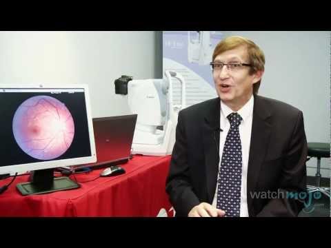
good okay so what are we looking at here
well everything is done we’re looking at
your fondest this is the back of the art
the pattern that you see is more precise
than a fingerprint we’re looking at
arteries and we’re looking at veins and
we’re looking at these white lines those
would be nerve fiber layer so we’re
talking about glaucoma so your cup to
disc ratio is perfect your artery vein
ratio is also very good and we’re then
going to look at your macula your macula
is your Center vision and this allows
you to actually see color and then it
allows you to see black and white so
when we separate the images
electronically we’re going to talk about
glaucoma and in your case York up to
this ratio is perfect your nerve fiber
layers are perfect so we are ruling out
in this instance the glaucoma aspect
then we’re looking at diabetic
retinopathy and we would see bleeders
small hemorrhages and we of course seen
on and then we’re going to look at deep
inside your choroid and these would be
for age-related macular degeneration
which you’re too young to have but these
are the tests so we’re looking for
glaucoma diabetes and age-related
macular degeneration and this gives you
a clean bill of health okay so there was
that second really bright flash so
what’s that photo yes the bright
photograph is a monochromatic view so
it’s a black and white picture we’re
looking into the layers of the RPE
called the retinal pigment epithelium so
in your case you’re extremely healthy
everything is fine and what we would do
in case you weren’t well from a
optometry point of view combined with a
nutrition we would change how you eat
what you eat your supplementation and
vitamins and so and so forth and then we
could change
the event of having an adverse vision
problem great thank you very much thank
you for your time
Please follow and like us: